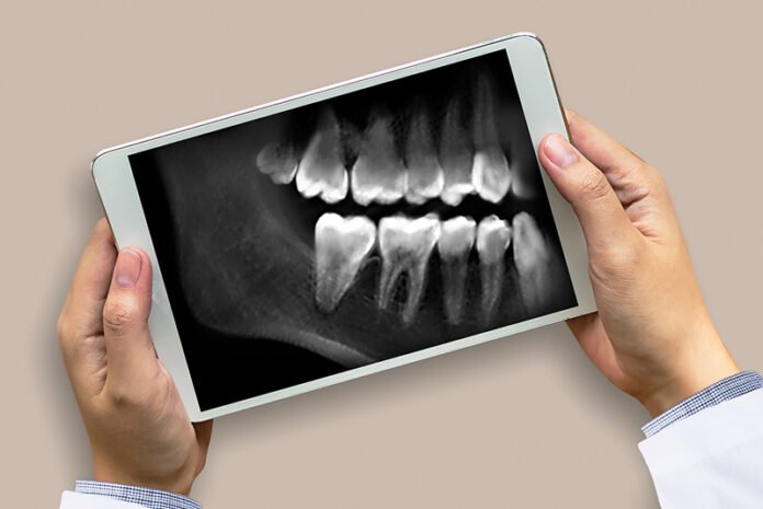
Everyone who has visited the dentist has had dental X-rays taken at some point, whether as part of a normal appointment or to aid in the diagnosis of a problem. So, what are dental X-rays used for, and why are they such a vital preventative and diagnostic tool for dentists?
There are several forms of dental X-rays, each serving a distinct purpose. These are the most frequent dental X-rays and the data they supply your dentist.
How Are X-Rays Conducted?
X-rays of your teeth are taken while you are sitting straight in a chair.
A lead apron will be draped over your chest and a thyroid collar will be wrapped around your neck by the dental professional.
For the photograph, the X-ray sensor or film will be inserted in your mouth.
When it comes to X-rays, most individuals have no issues or pain. The size and position of the sensor installation influence how comfortable or uncomfortable you are. The size of your mouth is also a consideration because a tiny mouth makes positioning the sensor more difficult. Taking X-rays should never be unpleasant; at worst, it should be awkward or uncomfortable.
If you have a sensitive gag reflex, you should inform your dental technician ahead of time. They can do things to assist keep the gag reflex at bay while having the X-ray. Children are particularly prone to gag reflexes and have difficulty with dental X-rays.
X-Rays of Bitewing
Bitewings are normally taken once a year (or as directed by your dentist) to aid in the detection of dental cavities between your teeth and to assess the bone level that houses your teeth.
X-rays of the periphery
This form of X-ray, also known as a PA, captures a whole tooth image from the very top of the tooth (crown) to the very tip of the root. Periapical X-rays are typically obtained if you are experiencing symptoms related to a specific tooth or as a follow-up to a surgery. If there is an abscess, anomalies in the surrounding bone structure, or extensive decay, your dentist can assist.
X-Rays of the Occlusa
These specialty X-rays are not as commonly utilised as the others, but they can give extremely useful information. They are often used to examine the roof or floor of the mouth for additional teeth, impacted teeth, abnormalities, jaw problems, and solid growths such as malignancies.
X-Ray Panoramics
A panorex X-ray is usually performed every 3-5 years (or whenever your dentist recommends), but it can also be taken by your orthodontic in preparation for braces and by an actual doctor in preparation for surgery, such as wisdom tooth extraction.
X-Rays on Digital vs. Plain Film
Because of its simplicity of use, efficiency, and, most crucially, the reduced quantity of radiation required and utilised, digital X-rays are beginning to replace traditional plain film X-rays. Digital X-rays are obtained using a specialised sensor that transmits the picture straight to a computer, where it may be examined immediately. If the picture from a plain film X-ray is too bright or too dark, it must be retaken. With a digital X-ray, you may alter the image on the computer to make it simpler to read. The dentist can also use the X-ray to zoom in on certain sections of the picture and even produce a bigger image.
